Home
>
Surgical Atlas
>
ORIF ZMC

ORIF ZMC

Open Dropdown

Preoperative Considerations

Consent:

  • Need for re-operation (eg. hardware failure, non-union)
  • infection
  • Loss of teeth
  • Entropion (retraction of lower lid causing irritation from lower eyelashes)
  • Facial scarring
  • Enopthalms and/or lagopthalmos

Anesthesia/Positioning:

  • Supine
  • OK for oral airway
  • OK for paralytics
  • Potential for oculocardiac reflex w forced duction testing & globe manipulation

Other:

  • None

Armamentarium:

  • Local anesthetic w epinephrine
  • Lacrilube
  • Corneal shield
  • 15 blade
  • Retractors (eg. toe ins, toe outs, army navy, minnesota) 
  • Daymar retractors
  • Jay freer
  • Malleable (small, medium, large)
  • Bovie (Set at 15 mixed for work around orbit)
  • Bipolar (Set at 20)
  • Debakey
  • Tissue pickups
  • Metzenbaums
  • #9 periosteal elevator
  • Midface plating system
  • Needle drivers
  • Caroll-girard t-bar and screw
  • Sutures (3-0 chromic gut, 4-0 vicryl, 5-0 plain gut and/or 5-0 prolene) 
  • Bacitracin

Technique

Injection of local anesthesia w epinephrine
Injection of LA w epinephrine to upper blepheroplasty site
Daymar retractor used to provide tension a retract lower lid. Malleable used to retract orbit. Bovie at 15/15 W used to outline transconjunctival incision.
Inciion through conjunctiva
Sounding for bone w movie and then taking incision down to inferior orbita rim
Attempt to stay pre-septal however often will go post-septal as seen with herniation of orbital fat
Bovie any tissue bleeders in the process
Blunt dissectonto expose fracture and inferior orbital rim
Fracture site exposed and noted to be mobile
Inferior orbital rim fracture reduced. Note th tetrapod zygoma may not be reduced entirely.
Replace corneal shield
Injection of LA w epinephrine. Incision was marked in a crease prior to the addition of LA which can distort the tissue
Incision w #15 blade through skin and subcutaneous tissue
Gentle blunt dissection through tissue layers to expose frontozygomaic fracture site
Exposure frontozygomatic fracture
Gentle currettage of the fracture site
Vestibular incision with bovie (15/15 W) staying 5 mm above the mucogingival junction
Incision extended down to bone
#9 subperioteal elevator used to create a full thickness mucoperiosteal flap and exposing fracture sites. Note caution when working around V2 branch of CN V
Fracture sites exposed
Midface plate pplied to frontozygomtic fracture site. Note the plate is fixated w monocortical fixation to the mobile segment to allow for manipulation and reduction.
Stab incision w 15 blade for Caroll-Girard screw placement at the malar prominence
Carolll-Girard screw and T bar placement allowing for manipulation an reduction of ZMC
Once the ZMC is adequately reduced as seen from alll views the remaining monocrtical screws are applied to the zygomaticofrontal fractue site. The zygomaticosphenoid (ZS) suture is the most important suture to note allignment and can be visualized throught the lateral brow incision.
The inferior orbital rim fracture is then plated with a long splanning midface plate while the bony segments are stabilzed.
Long spanning midface plate at the site of the zygomaticomaxillary fracture sites
Confiming adequate reduction at all plated sites
Closure with 3-0 chromic gut in a running fashion
Skin incision closure in al ayered fashion with 4-0 vicryl deeps and 5-0 plain gut vs 5-0 prolene. The subconjunctival incision can be closed with 5-0 chomic gut single interrupted sutures or left as is.
Note before leaving OR perform forced duction test to ensure no globe entrapment

Postoperative Considerations

Immediate:

  • Sleep on non-affected side during healing period (6-8 weeks)
  • Maintain immaculate oral hygiene
  • Bacitracin to skin incisions tid for 3 days

Follow Up:

  • Evaluate skin and intramural incision healing
  • Evaluate for facial symmetry as edema resolves. Evaluate for evidence of enopthalmos or lagopthalmos
  • Diplopia is not unexpected post op. Monitor and document for resolution.

Operative Note

Surgeon(s): ***

Assistant Surgeon(s):  ***

Preoperative Diagnosis: ***

Postoperative Diagnosis: Same

Procedure(s): ***

Anesthesia: General

Implants: None

Specimen:***

Drains: None

Fluids: See anesthesia record

EBL: Minimal

Complications: None

Counts:  Correct x2

Indications: ​***

Findings: As expected

Procedure in Detail:

The patient was seen in the preoperative holding area with a H&P was updated, consents were verified, surgical site marked, and all questions and concerns related to the proposed procedure were discussed in detail.  The patient was transferred to the operating room by the anesthesia team.  The patient underwent general anesthesia with endotracheal intubation. Tegaderms were placed over the eyes. The patient was prepped and draped in the standard fashion for maxillofacial procedures.  A time-out was performed and the procedure began.

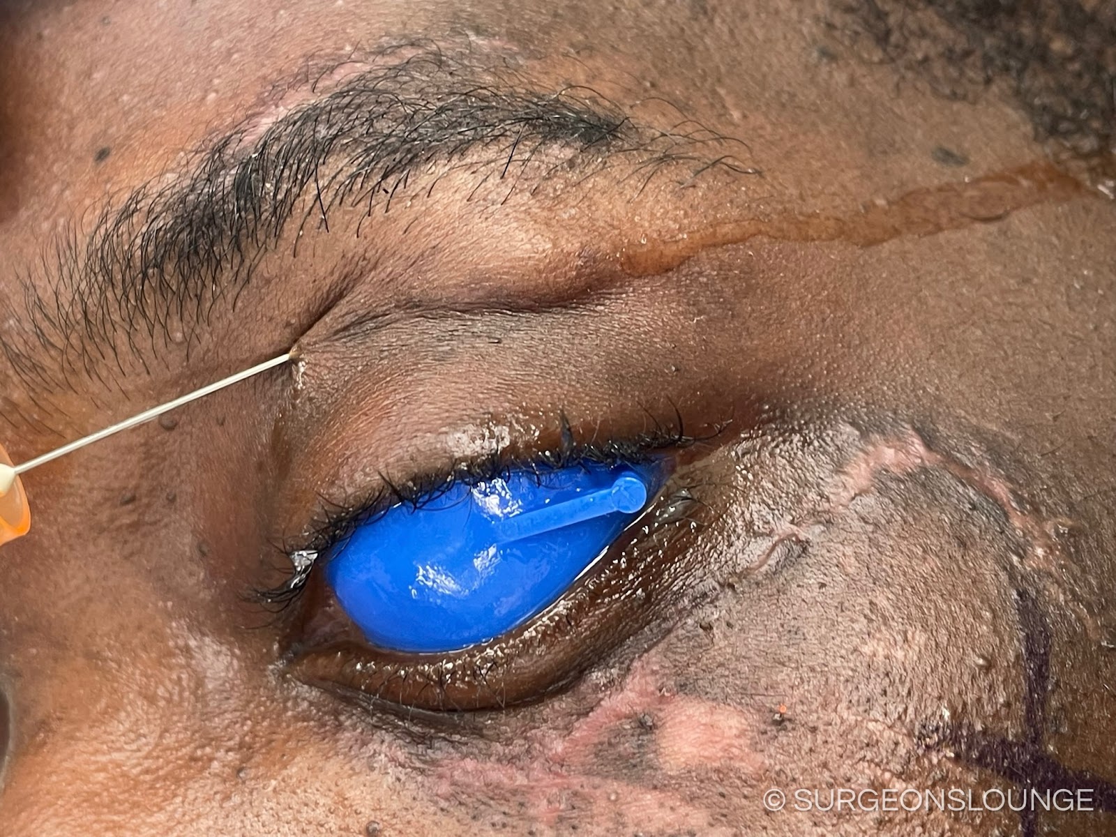
A forced auction test was performed with tissue forceps on the affected side noting full range of motion of the orbit. A corneal shield with lacrilube was placed over the globe. A marking pen was used to mark the site of the lateral brow incision. 10 ml of 2% lidocaine w 1:100k epinephrine was injected at the site of the lateral brow , transconjunctival, and vestibular incisions.

First attention was directed at the transconjunctival incision. A daymar retractor was used to retract the lower lid and provide tension while a malleable was used to retract the orbit. A bovie set at 15/15 W was used to make the initial incision through the conjunctiva. The bovie tip was then used to sound to bone and the movie was used to deepen the incision to the infraorbital rim. A #9 subperiosteal elevator and blunt dissection was used to expose the infraorbital rim and the fracture. The fracture was gently curettage and derided.

Next attention was directed to the lateral brow incision. A 15 blade was used to make an incision through skin and subcutaneous tissue. Next blunt dissection was used to go down to bone and the fracture site was exposed.

Next attention was directed intramurally. An incision was made in the maxillary vestibule with a bovie staying 5 mm away from the mucogingival junction. A #9 subperiosteal elevator and blunt dissection were used to bluntly dissect to to the site of the fracture being conscious not to damage the infraorbital nerve.

Attention was then directed to the fracture site at the frontozygomatic suture. A 4 hole mid face plate was placed at the site of the fracture and the plate was stabilized on the stable skull base side. A 15 blade was used to make a nick incision at the molar prominence and a Caroll-Girard screw was placed in stable bone a the zygoma and manipulated to an ideal reduction. The plate over the frontozygomatic suture was then fixated with mono cortical fixation.

Next attentio was directed at the inferior orbital rim where a good reduction was noted. A mid face plate and mono cortical screws were used to fixate the fracture site. Next attention was directed intramurally and a long spanning mid face plate was used to span the fracture and was fixated using mono cortical screws under copious irrigation.

The corneal shield was removed and a forced duction test was performed showing full range of motion of the globe. Closure of the skin incisions was completed in a layered fashion using 4-0 vicryl and 5-0. The corneal shield as replaced and the conjunctival incision was closed with 5-0 chromic gut single interrupted sutures. The vestibular incision was closed with 3-0 chromic gut in a running fashion. The globe was irrigated with ophthalmic solution.

The patient's face was then cleaned and the posterior pharynx was suctioned. Bacitracin was placed over the skin incisions. An OG tube was used to suction out the contents of the stomach. Tegaderms were removed from the eyes. Dressings were placed. The patient was then transferred back to the care of the anesthesia team for extubation and recovery.

Coding

  • 21365 ZMC through multiple approaches

Comments